The eyepiece and an objective lens. Light microscopes magnify the image of the specimen using light and lenses.

The Parts Of A Compound Microscope And How To Handle Them Correctly Human Anatomy And Physiology Lab Bsb 141
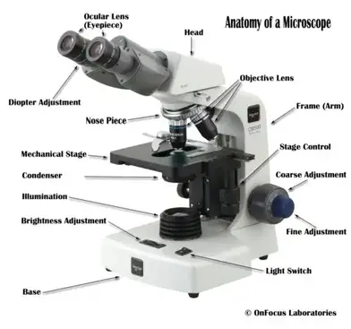
The entire microscope is handled by a strong and curved structure known as the arm.
. In a compound microscope the sample is illuminated from the bottom to observe transmitted light or from the top to observe reflected light. The compound microscope is mainly used for studying the structural details of cell tissue or sections of organs. The stage is the flat platform where the slide is placed.
Parts of Compound Microscope. Nosepiece is a rotating turret that holds the objective lenses. 1tissues 2organ systems 3organelles 4organs.
Always place the microscope on a level and stable surface. When you use a compound light. This type of microscope might be used to study external features on an object.
Light microscopes _____ the image of the specimen using _____ and _____. The viewer spins the nosepiece to select different objective lenses. The base is also known as the foot which is either U or horseshoe-shaped.
Microscope slides should always be prepared with a cover slip or cover glass over the specimen. It is a U-shaped structure and supports the entire weight of the compound microscope. Grasp the arm with one hand and place the other hand under the base for support.
- Animal cells have centrioles for cell division. The objective and the eyepiece whose individual powers multiply to enable much higher magnifications than those achieved by a. A compound light microscope has its own light source in its base.
A dissecting microscope is used to view three-dimensional objects and larger specimens with a maximum magnification of 100x. The illuminator is the light source for a microscope. The lens closest to the specimen is called the objective lens while the lens nearest to the users eye is called the ocular lens or eyepiece.
Which structure is best observed using a compound light microscope. It is a vertical projection. The compound microscope uses two lenses to magnify the specimen.
This stands by resting on the base and supports the stage. - Plant cells have cell walls and chloroplasts. 6Which structure is best observed using a compound light microscope.
1organs organism cells tissues 2organism cells organs tissues 3cells tissues organs organism 4organism organs tissues cells 7Which sequence shows a decreasing level of complexity. There are two pairs of screws for moving the body tube in relation to stage larger for coarse adjustment and. By soetrust February 4 2022.
Therefore a 10x eyepiece used with a 40X objective lens will produce a magnification of 400X. The parts of a compound microscope can be classified into two. The term _____ means that this microscope passes through light through the specimen and then through two different.
This will help protect the objective lenses if they touch the slide. A light microscope might be used when examining individual cells within living tissue. O the inner surface of a mitochondrion O a cell O a DNA sequence O a virus.
The microscope has a rack and pinion attached either to body tube or the stage for bringing the object under focus. Light from the sample is collected by an optical system consisting of two main lens groups. A compound light microscope mostly uses a low voltage bulb as an illuminator.
Differences between plant and animal cells. Magnify light lenses. The term compound means that this microscope passes light through the specimen and then through two different lenses.
- Animal cells have many small vacuoles while plant cells have big vacuole. Microscopes are used to study thing that are too _____ to be easily observed by other methods. Cell structure that directly controls regulates the movement of molecules into and out of the cell.
The incandescent light from the light source is reflected by a condenser lens beneath the specimen and the light passes through the specimen up to the objective lens then the projector lens sends the magnified image onto the eyepiece. In order to ascertain the total magnification when viewing an image with a compound light microscope take the power of the objective lens which is at 4x 10x or 40x and multiply it by the power of the eyepiece which is typically 10x. In most microscopes there is a choice of objectives to use.
It typically has a magnification power of up to 1000x.

Parts Of A Microscope With Functions And Labeled Diagram
Microscope Parts And Functions

What Is A Compound Microscope New York Microscope Company
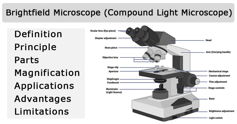
Brightfield Microscope Compound Light Microscope Definition Principle Parts


0 Comments